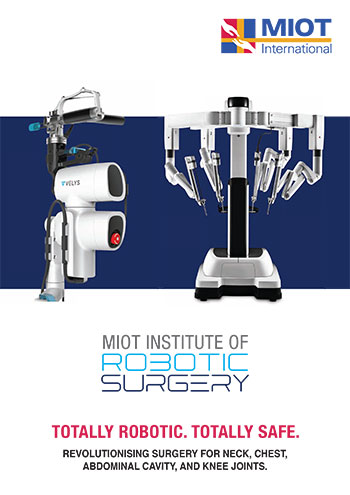


MIOT Institute of Robotic Surgery 2
TOTALLY ROBOTIC. TOTALLY SAFE.
REVOLUTIONISING SURGERY FOR NECK, CHEST,
ABDOMINAL CAVITY, AND KNEE JOINTS.
MIOT’s Cutting-Edge Robotic Surgery
- Safer surgery with high precision
- Low healthcare costs
- No complications
- Faster recovery times
- Better patient outcomes
- Minimal pain
- Reduced blood loss
- No scar
- Shorter hospital stay
- A quicker return to quality of life
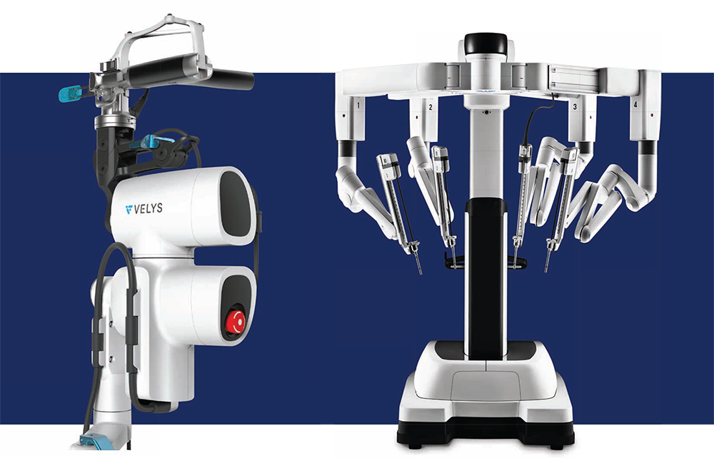
MIOT is dedicated to integrating the latest advancements in medical science to provide the best care for patients and enhance outcomes. Understanding the need for cutting-edge technology in today’s world, we have launched the MIOT Institute of Robotic Surgery. The Da Vinci Xi Robotic Surgical System and Robotic-Assisted solution for Total Knee Replacement were introduced at the right time of their development to enhance MIOT’s medical technology and improve patient outcomes. The Da Vinci Xi Robotic Surgical System is designed to perform general surgeries from the neck to the abdomen, whereas the Robotic-Assisted Total Knee Replacement is specifically used for total knee replacement surgeries.
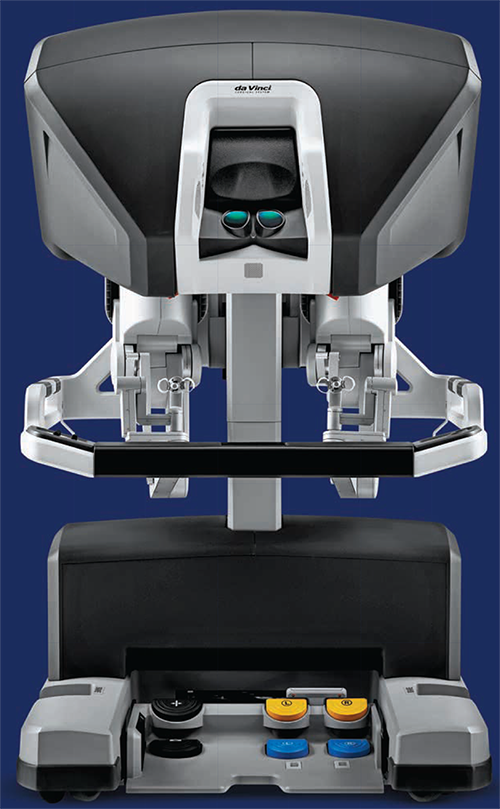
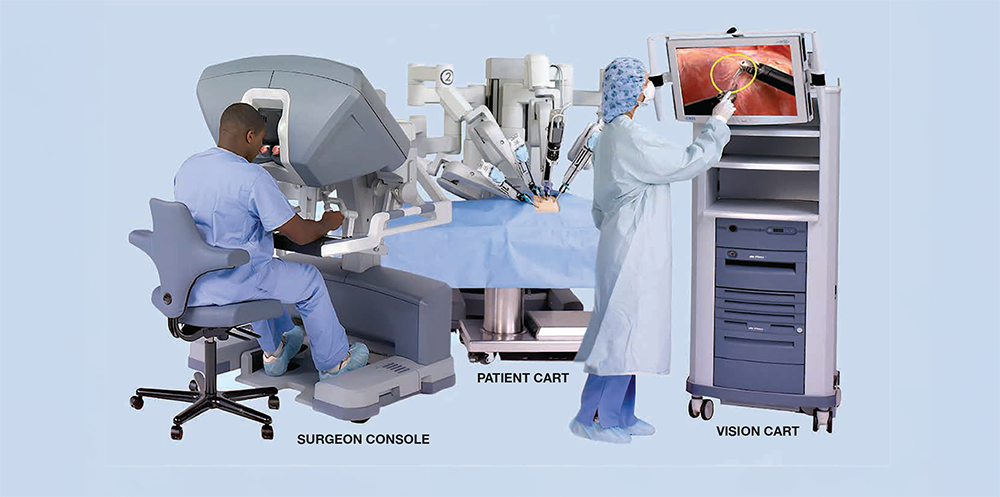
Da Vinci Xi Robotic Surgical System
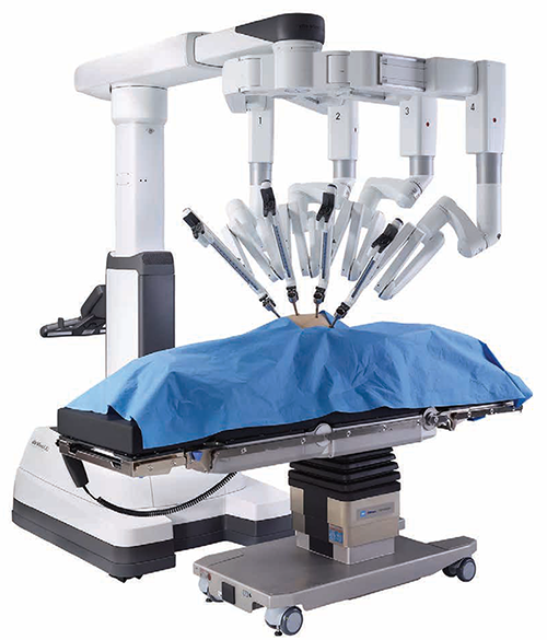
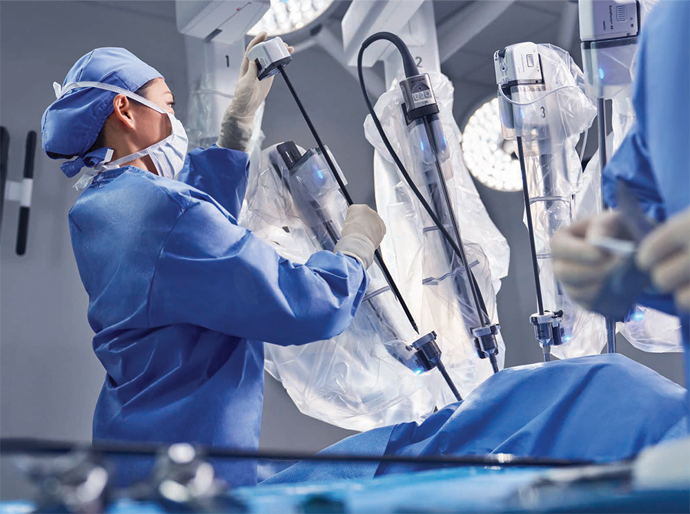
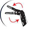
Robotic surgery is an advanced form of minimally invasive or laparoscopic (small incision) surgery in which surgeons perform the surgery using a computer-controlled robot, with the surgeon having complete control of the robotic system. This technology allows complex surgeries that require high precision to be performed safely without damaging surrounding tissues or organs. Robotic technology enhances precision, minimises surgical complications, and improves patient outcomes. This technology ensures a high level of safety for patients undergoing over 140 different types of surgeries. The enhanced safety of patients is further increased by the Robotics-Assisted Intraoperative Ultrasound System. The transducer of this advanced ultrasound system, placed inside the body, provides high-resolution images with great anatomical details during surgery. These real-time images are captured from all angles with full robotic articulation. It provides greater accuracy and precision to ensure the identification of the entire tumour surface for complete tumour resection.
Concerning Factors of Surgery
No More Fear of Surgery: People often delay surgery due to concerns about safety, recovery, and regaining quality of life. This may worsen their condition and eventually lead to critical health issues. However, these fears can now be alleviated with the use of Robotic Surgery.
Robotic technology provides a great comfort for the surgeons. The surgeon sits in the surgeon console and performs the surgery. This eliminates their fatigue and improves their performance. This not only reduces the risk of errors but also improves overall patient outcomes, making surgery safer and more effective.
The advancements include


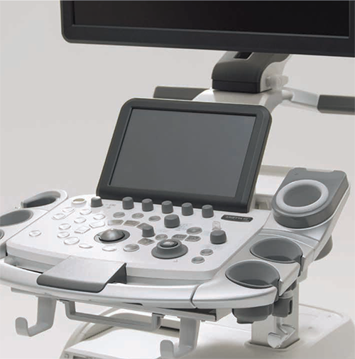
Robotics-assisted intraoperative ultrasound system The success of surgery largely depends on an accurate diagnosis. Diagnostic imaging is typically performed a day or even a week before surgery. The actual size or depth of a tumour or other abnormality may change by the time of the procedure, potentially impacting the surgical outcome. This advanced Robotics-Assisted Intraoperative Ultrasound System aids surgeons by allowing them to locate and accurately visualise anatomical abnormalities in real-time during surgery. It helps precisely identify the tumour’s location, depth, and borders, making it easier to distinguish between the tumour and normal tissue.