
Past events at MIOT
MIOT International Successfully Treats a 46-Year-Old Mauritian for a Rare and Potentially Catastrophic Case of a Foreign Body in the Right Side of the Neck.
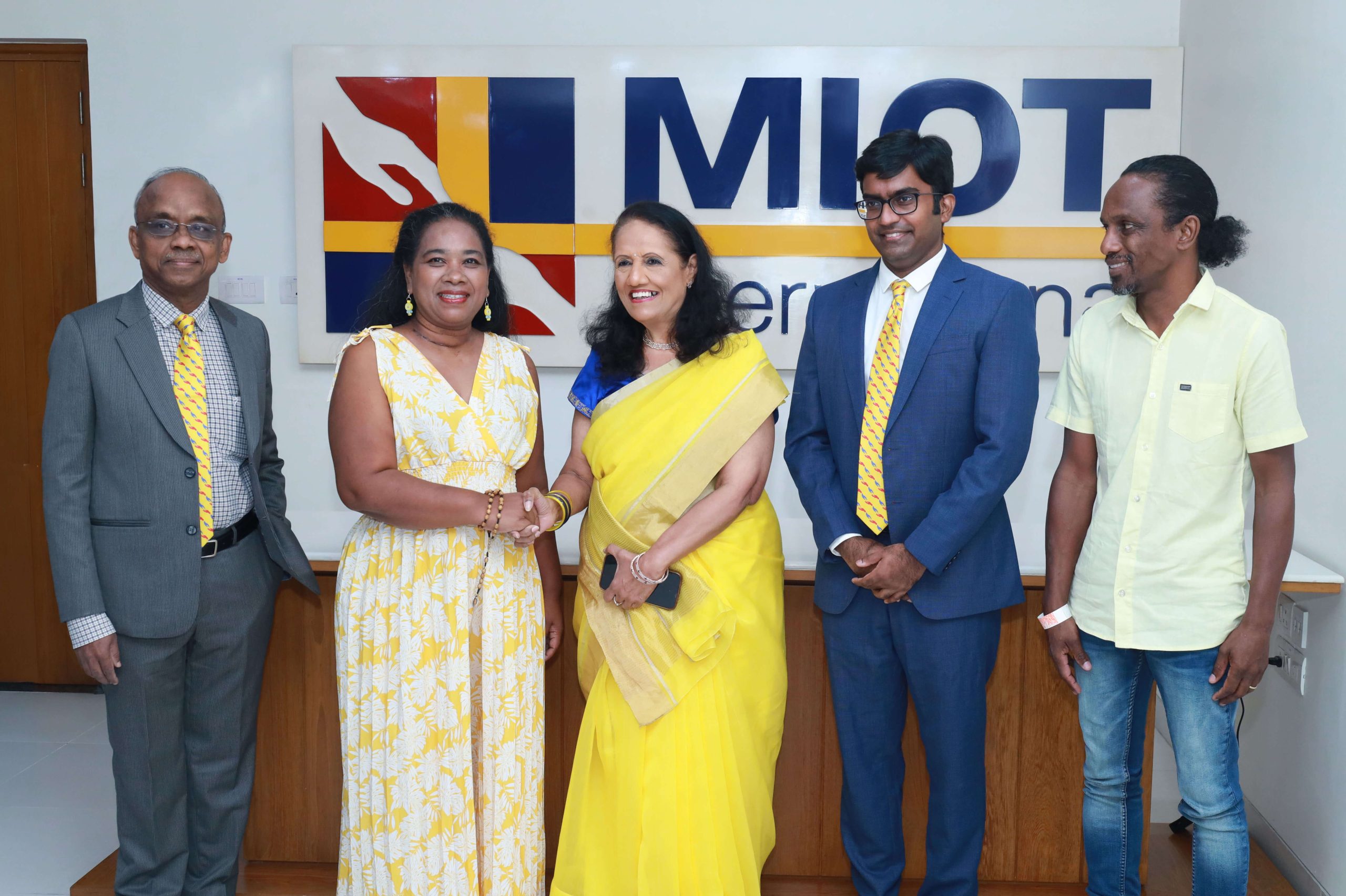
From Left to Right:
Dr.Palaniappan S, Senior Medical Gastroenterologist, Hepatologist & Interventional Endoscopist, MIOT International;
Ms. Marie Desiree Isabelle Heliotrope, Mauritian Patient who successfully underwent surgery at MIOT International;
Mrs. Mallika Mohandas, Chairman, MIOT International;
Dr. Arun Mitra Singamaneni, Head & Neck, Thoracic Onco Surgeon, MIOT International;
Mr. Teddy Emmanuel Berty Heliotrope, Husband of patient.
India has emerged as a leading global destination for advanced medical care, with MIOT International playing a major role in making Chennai a top destination for medical excellence. The institution offers cutting-edge technology and multidisciplinary expertise to promptly manage critically challenging cases.
Clinical Presentation and Initial Evaluation: A Painful Ordeal with No Clear Answers
A teacher and a mother of three, Mrs. Marie Desiree Isabelle Heliotrope, a 46-year-old woman from Mauritius, began experiencing severe throat pain and difficulty swallowing after eating a meal on 24th April 2026.
What seemed like a minor discomfort soon turned into a frightening and painful ordeal. As the pain worsened with each passing day, even eating and swallowing became extremely difficult. She went from hospital to hospital in Mauritius searching for answers. Doctors performed scans and passed a camera into the food pipe, identifying a foreign body in a critical region of the neck, leaving doctors uncertain about how it had migrated there. With no relief in sight, she was left exhausted, anxious, unable to eat properly, and in constant pain.
The doctors in Mauritius advised her to urgently travel overseas to a hospital with multidisciplinary expertise, which was not available locally. Desperate for a solution, she reached out to her international insurance provider for urgent medical assistance.
Although treatment options were available across Europe, North America, and South Africa, India was chosen. This decision reflects the increasing global trust in Chennai’s exemplary medical expertise and its world-class healthcare infrastructure, making India not just a viable option but a premier destination for advanced medical treatment.
Foreign Body Migrated from the Food Pipe into the Neck
On arrival, a detailed evaluation was conducted at MIOT International without any delay. The evaluation revealed that a 24 mm foreign body had pierced through the food pipe and migrated into a highly critical region of the neck.
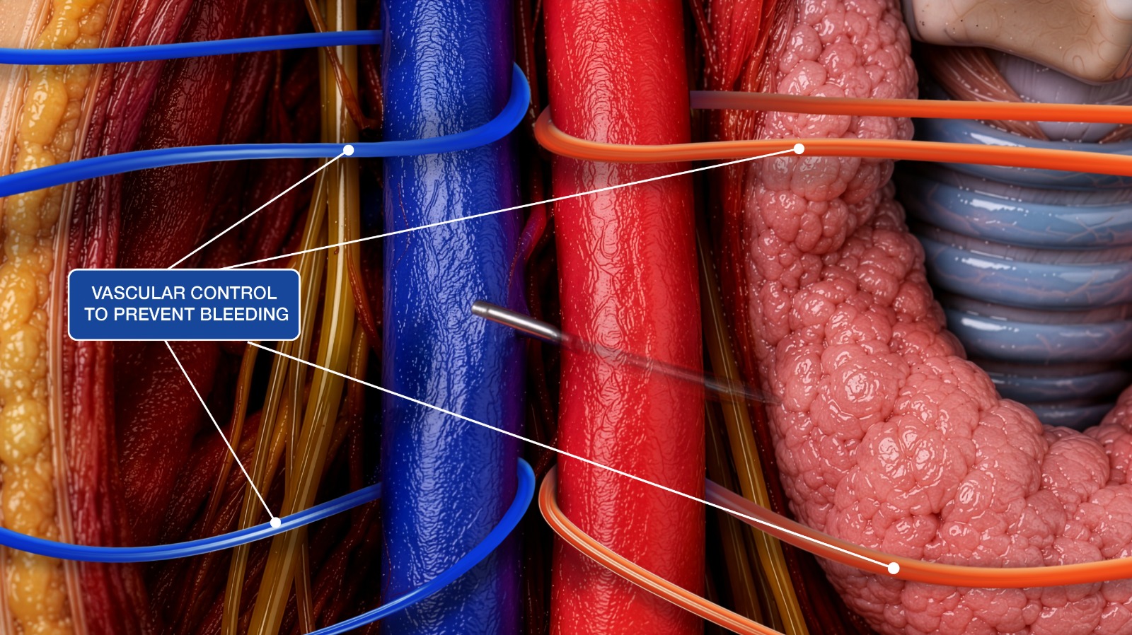
The foreign body had penetrated between two major blood vessels, the right common carotid artery and the internal jugular vein. The right common carotid artery carries oxygenated blood from the heart to the head and neck, while the internal jugular vein carries deoxygenated blood back from the head and neck to the heart.
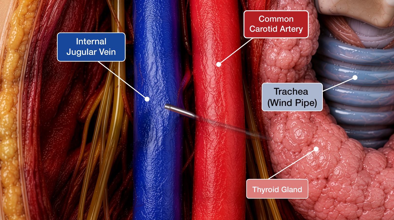
The foreign body was also found in proximity to the windpipe (trachea) and the thyroid gland, making the condition highly complex and potentially life-threatening.
Any sudden movement or imprecise surgical handling could have resulted in catastrophic bleeding, airway compromise, cardiac or neuro-related complications.
Where Every Step Mattered
One of the biggest challenges in such cases is that the foreign bodies can shift position once the patient is anaesthetised and positioned on the operating table.
In many situations, surgeons may spend hours searching for the foreign body while navigating around delicate structures. However, in this case, extensive exploration was not possible because the foreign body was surrounded by delicate, vital structures, including the artery, veins, windpipe, and thyroid gland.
Intraoperative Findings and Surgical Management
Given the complexity and potentially life-threatening nature of the case, a multidisciplinary team comprising specialists from Head & Neck Surgery, ENT, Medical Gastroenterology, Anaesthesiology, and Radiology came together to plan the treatment approach. Their collaborative expertise played a crucial role in ensuring accurate diagnosis and safe surgical management.
Before the surgery, in the operating theatre, an endoscopic evaluation performed by a medical gastroenterologist confirmed that the foreign body had already punctured the food pipe and migrated toward the right side of the neck.
To further overcome the challenge of locating the foreign body in such a delicate area, the surgical team utilised an advanced intraoperative CT imaging system (BodyTom), which enabled real-time pinpointing of the foreign body after the patient was positioned for surgery.
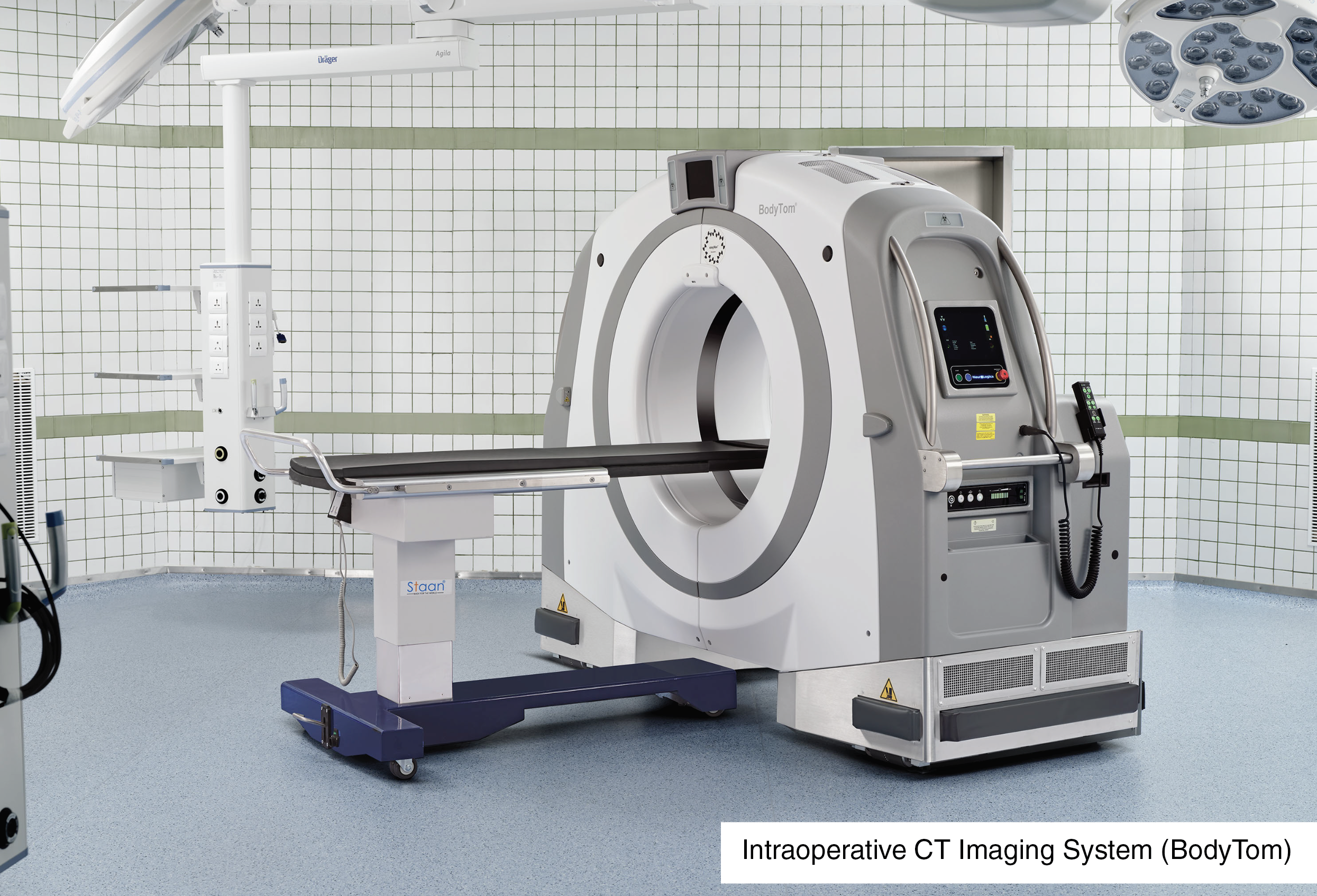
The continuous real-time monitoring throughout the procedure allowed the team to accurately identify the exact location of the foreign body and avoid unnecessary damage to critical surrounding structures.
While making the incision to remove the foreign body, the ENT and Head and Neck surgical team observed significant inflammation in the area. To the team’s surprise, the foreign body was a 24 mm metallic wire lodged between the major blood vessels and adjacent vital structures.
Precision Surgery Performed Without Damage to Vital Structures
Given the high-risk location, vascular control measures were immediately established to prevent bleeding before the metallic wire was carefully removed.
The procedure was performed with extreme precision while preserving all surrounding vital structures. Intraoperative CT imaging confirmed complete removal of the foreign body with no metallic residue, vascular injury, or airway compromise.
After meticulous extraction of the foreign body, the puncture sites were secured to prevent bleeding from the blood vessels.
Successful Recovery and Restoration of Quality of Life
The post-operative period was uneventful. The patient remained stable and was continued on supportive care.
After 2 days post-surgery, oral liquid feeding was initiated, and imaging was performed to confirm that there were no leaks.
Within a week, she was able to eat solid food comfortably again and was relieved to regain her normal swallowing and voice without any discomfort or pain.
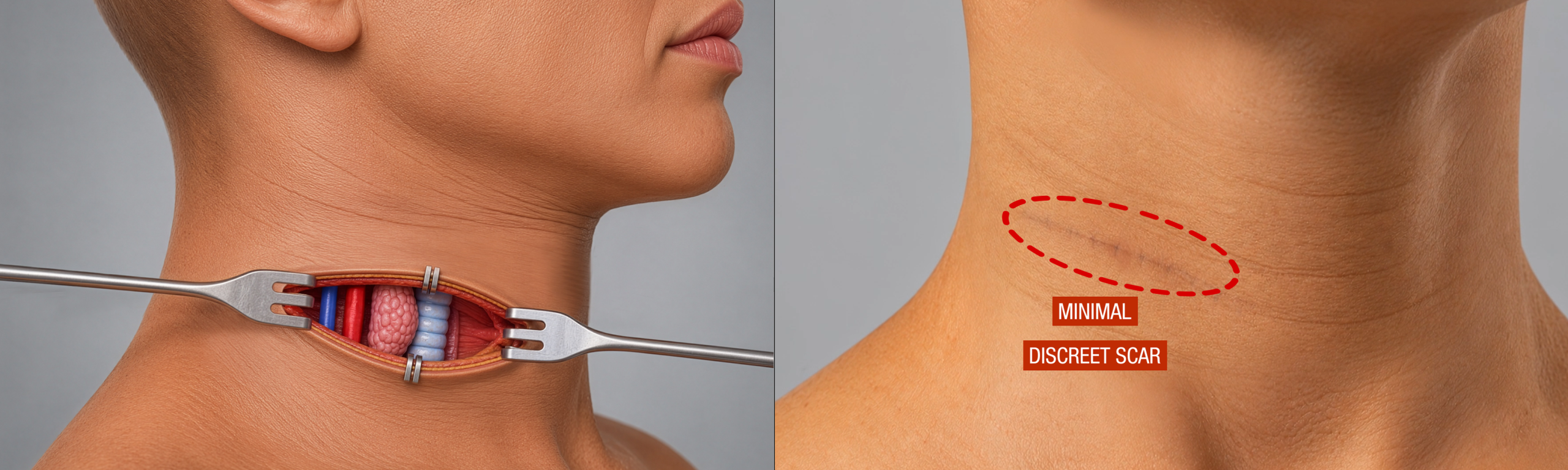
The incision was made in a way that the scar is discreetly concealed within the natural crease of the neck, where it is expected to continue fading over time. This approach not only provides an excellent cosmetic outcome but also benefits the patient by building their confidence and self-esteem.
India’s Surge as a Leading Global Healthcare Destination
This case reinforces India’s growing position as a trusted destination for advanced and complex medical care, with patients increasingly travelling to Chennai when timely and specialised treatment is critical.
In most cases, the patient’s swift transfer from their country to Chennai plays a major role in ensuring they receive the right care without any delay. In such emergencies, even a few hours can significantly alter outcomes.
MIOT International, Chennai, today offers a unique combination of state-of-the-art medical technology and the rigorously trained expertise of highly experienced specialists. This enables precise diagnosis and advanced surgical care in the most complex situations. It is further strengthened by cost-effective treatment, making world-class healthcare accessible to patients from across the globe.
The availability of direct flights could be a crucial factor in preventing potentially life-threatening complications and ensuring the patient reaches expert care at the right time, when precision and speed matter the most.
This case once again highlights a simple truth in critical medical situations: “Time is life”.
As Chennai continues to emerge as a global healthcare hub, direct international connectivity will play an important role in enabling faster access to specialised treatment and saving lives from all around the world.