Past events at MIOT
For the 1st time in India, MIOT Hospitals brings a Biplane CathLab with Cone Beam CT, 3D Echo and software intelligence – all on a single platform
INTRODUCING MIOT HOLISTIC INTERVENTIONAL SUITE
An institution built on innovation, MIOT Hospitals has always been at the forefront, embracing the latest in healthcare technology to revolutionize patient care in India. It was a watershed moment in Indian orthopaedics when MIOT pioneered Computer-Navigated joint replacement in Asia Pacific. Cancer patients gained the confidence to defeat tumours when MIOT introduced image-guided radiation therapy for the 1st time.
From conceiving pinhole surgery to unveiling the state-of-the-art 3T Silent MRI machine, technology is at the heart of everything we do.
A new era in Indian healthcare begins today as we bring patients a futuristic machine that is about to take the healthcare world by storm. MIOT Hospitals redefines your future healthcare with the holistic Interventional Suite.
For the 1st time in India, MIOT brings together a Biplane CathLab System with a Cone Beam CT and a 3D Echo machine, supported by software intelligence.
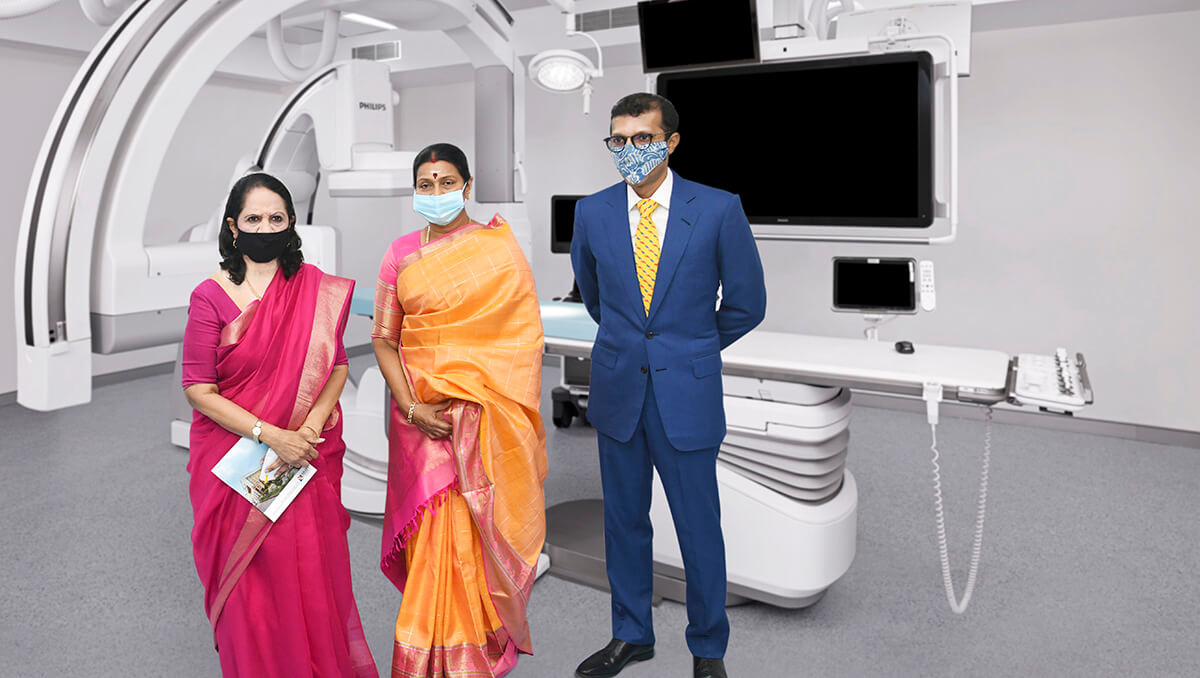
Smt. Durga Stalin inaugurated the Holistic Interventional Suite (27th August 2021) at MIOT Hospitals . Seen along with is Mrs. Mallika Mohandas, Chairman, MIOT International and Dr. Prithvi Mohandas, Managing Director, MIOT International.
MIOT INTERVENTIONAL SUITE – THE FUTURE IS HERE
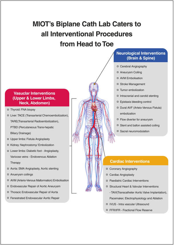
Two C-Arms
MIOT Interventional Suite’s Biplane CathLab has two C-arms as opposed to just a single arm in the conventional system. The Biplane’s 2 C-arms integrate diagnosis, on-the-spot decision-making and treatment on a single platform to save precious time. Multiple views can be obtained in a single move. This reduces contrast usage and exposure to radiation by manifold.
Its in-built Cone Beam CT gives specialists the rare advantage of providing CT-like images on the table within a few seconds. The 56’’ LCD screen that comes with the system gives a clear 3D picture of the organs in question, so that margins of error are brought down to zero.
Single Platform Advantage
The Biplane CathLab integrates diagnosis, decision making and treatment on a single platform. Hence, patients need not be shifted from the CT room for diagnosis to the treatment room and back. Moreover, by having the patient for diagnosis and treatment in the same room, critical decisions are taken on-table without loss of time.
These attributes make the system superior by manifolds than the conventional CathLab system.
Benefits of the MIOT Biplane CathLab System:
SPEED
Speed is a crucial factor when it comes to stroke. Patients brought in with symptoms of stroke have to be treated quickly to save maximum brain tissue and to avoid irreversible brain damage.
Stroke
- The MIOT Biplane CathLab System significantly brings down the treatment time for a stroke patient from 2 hours to a mere 30 minutes. This is by far a superior milestone that means patients can rest assured of being saved without much complexity.
- Conventionally, to identify whether stroke is due to a block or a bleed, it takes 20 minutes with a normal CT scan. The patient then needs to be shifted to CathLab for treatment.
- But now, patients can be directly taken to MIOT’s state-of-the-art Biplane CathLab where the CT is taken by its in-built Cone Beam CT to captureCT-like images in just 5.2 seconds.
- In case the stroke is caused by blockage in the brain vessel, the exact location of the vessel block (the brain vessels take 360 loops and superimpose on each other) needs to be identified before planning the treatment process.
- Conventionally, this requires 12 Angiograms (12 contrasts and 12x radiation exposures) at various angles put together and delays the treatment.
- Now with MIOT’s state-of-the-art Biplane CathLab System, just 1 Angiogram (1 contrast and 1 Radiation exposure) is enough to obtain a complete 3D image of all the brain vessels using SMART CT ANGIO. This way, the exact location of the vessel block is identified within a few seconds and the treatment approach is planned in advance.
- Using the same images from Smart CT Angio, a 3D Roadmap is created using SMART CT ROADMAP application to navigate to the block and remove it or place a stent to dilate the vessels.
- Using SMART CT VASO application, the stent is placed well close enough to the vessel wall and is confirmed on the table itself without the need for moving the patient to CT SCAN again to confirm the same.
ACCURACY
Blood vessels are highly intricate structures and they demand high level of precision to perform complex procedures like valve placements. When the images of target sites are clear, specialists can reach damaged valves of the heart and perform complicated procedures with ease.
TAVI
Trans-catheter aortic valve implantation (TAVI) is a minimally invasive procedure in which a new valve is inserted without removing the old, damaged valve. This is done by inserting a catheter into a blood vessel in your upper leg or chest and passing it towards your aortic valve.
- Conventionally, cardiac CT- Angiogram is taken to identify any disease of the heart valves. The doctors would have to anatomically imagine (using old CT scan of the heart) and guide the catheter wire into the vessels. Many angiograms need to be done to confirm if the catheter is on the right track while reaching the aortic valve. Also 3D echo data and X-ray data on table requires spending valuable time and effort to mentally align them during the procedure for aortic valve replacement.
- But now with MIOT’s state-of-the-art Biplane CathLab, pre-acquired CT scan of the heart can be imported into the system and reconstructed as a 3D structure of the heart using HEART NAVIGATOR-TAVI application avoiding the need for contrast.
- Using 3D DYNAMIC ROADMAP – real-time, automatic, motion-compensated images help while deploying the artificial valve to the aorta.
- Ultrasound images captured from the EPIQ- CVxi, a 3D echo machine is fused / overlaid with live x-ray images taken in the MIOT Biplane CathLab machine using ECHO NAVIGATOR This helps in accurate placement of the valve in the aorta. This also plays an important role in dealing with structural heart disease especially in paediatric patients.
SAFETY
It’s not just accuracy and time, but the MIOT Biplane CathLab System’s unique features make it a leader when it comes to patient safety. The machine reduces the use of contrast dye by nearly 50%, ensuring safe treatment for patients with kidney and liver issues.
Moreover, the MIOT Biplane CathLab’s procedure card application uses patient history to customize radiation levels depending on the patient’s age and medical condition.
TACE
Trans-arterial Chemoembolization (TACE) is a specific procedure to deliver chemotherapy agents to cancer sites of the liver.
- Conventionally, healthy liver tissue along with cancerous tissue is also damaged in the process of chemoembolization of cancer cells. Moreover, multiple angiograms were taken to identify the exact vessels supplying the cancer site.
- With the new MIOT Biplane CathLab System’s CONE BEAM CT, entire soft tissue information and the vessels supplying tumour sites are identified in just a single angiogram.
- With the EMBO GUIDE application, 3D structure of the tumour and vessel supplying the tumour along with a 3D roadmap are obtained. This assists specialists to navigate the catheter into the target vessels and selectively deliver the chemo agents without compromising healthy liver tissues.
- Thus, patients are relieved of the undesirable side effects and can resume their normal lifestyle sooner than ever before. The use of contrast dye and the exposure to radiation are also brought down by 50%.
Redefining the future of healthcare and promising a high standard of care to patients have become a reality today. As MIOT launches a fully-configured futuristic Biplane CathLab System for the 1st Time in India today a new dawn is here in Indian healthcare.
| Conventional CathLab Vs. MIOT Biplane CathLab | ||
|---|---|---|
| Procedure | Conventional CathLab | MIOT Biplane CathLab |
| Stroke treatment | 2 hours | 30 minutes |
| Contrast Dye Usage | Minimum 70 ml | Maximum 20 ml |
| Radiation Exposure | X amount | Only 50% of X amount |
| CT Scan Time | Conventional CT Scan 20 mins | Near CT like images Just 5.2 sec |

72 yr old Lady eats without pain after 3years
A successful case performed at MIOT’s State-of-the-art Biplane Cath Lab
Mrs. Shanti, a 72 year old lady had a history of stomach pain for the past 3 years. She was admitted with complaints of severe abdominal pain on eating 1year ago. The pain was so extreme, that eating had become a curse for her. She survived on very minimal amounts of semi-solid and liquid food which had made her lose a considerable amount of weight.
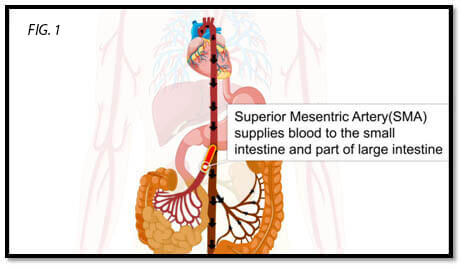
She was diagnosed to have (mesenteric ischemia) blockages of the two of the three main blood vessels supplying her intestines.
Considering her age she was treated with medicines for 6 months but her pain persisted and she started passing blood in her stools hence she got admitted at MIOT last month.
She was not fit for surgical treatment due to her age and the need for major high risk surgery to treat it. She was referred to our Interventional Radiologist to see if this could be treated by endovascular procedure.
Endovascular surgery is an innovative, less invasive procedure used to treat problems affecting the blood vessels that involves making a small incision near each hip to access the blood vessels.
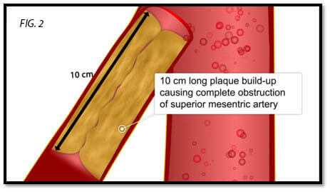
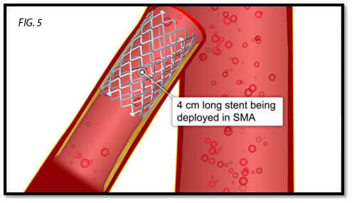
Patient had a 10cm long standing hardened blockage of the main Superior Mesenteric Artery (SMA) (Fig. 1 & 2) and the chances of successful reopening were slim. But considering her age, pain intensity and blood loss, MIOT Doctors decided to take her up for the interventional procedure.
The next day she underwent the surgery in the MIOT’s State-of-the art Biplane Cath lab. As the block was in a very complex position, images were taken simultaneously in 2 projections using the 2 C-arm which was crucial to opening the completely blocked blood vessel in this case. In a machine with a single arm, this would have required the C-arm to be rotated frequently between the 2 required positions, leading to a longer procedure and also the risk of wire getting dislodged from crucial positions when the C-arm is being swung.
Also, with 2 C-arms, only 1 Angiogram is needed as both angulations are captured simultaneously with 1 Contrast dye injection and 1 Radiation exposure, but in conventional Cath lab, 2 Angiograms are needed every time, thus the New Biplane Cath lab reduces the contrast and radiation dose by 50%.
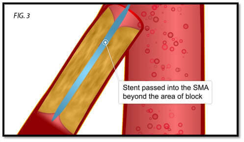
In MIOT’s state-of-the-art Biplane Cath Lab, SMART MASK application overlaps the Angiogram images on live X-ray images, used for live guidance of the catheter.
With the guidance of a SMART MASK application, the interventionist managed to open her blood vessel by slowly crossing the blocked segment (Fig. 3).
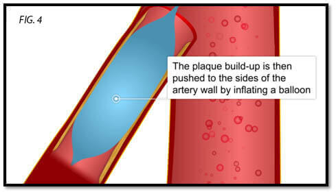
As the block was 10cm long, a balloon was inflated to push the plaque towards the walls of the vessels (Fig. 4).
Then a 4cm long stent was placed to keep the vessel open (Fig. 5).
The next day the patient’s pain had resolved and she enjoyed her morning cup of hot tea after a very long time. Slowly semisolid food was introduced and now she is managing it quite well. She also stopped having blood in the stools.