

Past events at MIOT
Successful Endovascular Stenting Performed 1ST IN INDIA on a 15 year-old-boy with extremely complicated Aortic Dissection Through a Hybrid procedure.

Seen in the photograph from left to right: Patient’s father, Mrs. Mallika Mohandas, Chairman – MIOT International, Patient (Mr. Bharath), Dr. Vijit K. Cherian, Director – Adult Cardiothoracic Surgery, MIOT International, Dr. K Murali, HOD – Interventional Radiology, MIOT International.
Mid May 2017, 15 year old Bharath was referred from Vizag, with an unusual diagnosis for a boy of his age. He was presented to MIOT with a diagnosis of COMPLICATED AORTIC DISSECTION in the descending aorta.
Earlier (April last week) when Bharath visited a local hospital at Vizag (after his regular swimming) with vague symptoms like fainting attack and unable to move his limbs, the doctors suspected Bharath to have some neurological issues. So he was treated for muscle disorder and was then discharged. But later when he came back with symptoms of severe pain in the abdomen, the doctor noticed that there was no pulsation to his lower limbs. A CT angio was performed and it showed that Bharath had Aortic Dissection.
The Vizag doctors referred Bharath to MIOT Hospitals immediately for further diagnosis and treatment. At this stage Bharath could hardly walk 10 steps and he was rushed to MIOT in an ambulance from the Chennai Airport.
Investigations at MIOT:
Further investigations at MIOT by the Cardio Thoracic Surgery team revealed that the AORTIC DISSECTION in the descending aorta was so extensive that most of his blood supply to the abdominal organs and the legs were severely compromised. The cause for such a dissection in this age group is not known and when it does occur, other cardiac defects are commonly seen. In Bharath’s case it is most likely due to a Connective Tissue Disorder without genetic linkage as nobody in his family had any related disorder.
The Cardio Thoracic Surgery team discussed Bharath’s case with the Endovascular team. Ideally surgical repair would be the 1st treatment option. However due to complexity of the case it was obvious that this case carries chances of high mortality and morbidity including the possibility of paraplegia (an impairment in motor or sensory function of the lower half of body). Hence the team came to a consensus that a Hybrid procedure was the most suitable option for this boy. Medical literature had very few cases of such a presentation.
On 23 May 17, the MIOT team of doctors under Dr. Vijit K Cherian (Director – Adult Cardio Thoracic Surgery) and Dr. K. Murali (HOD – Interventional Radiology), went ahead with a Hybrid procedure.
Hybrid Procedure:
Due to the small blood vessels and dissected arteries to his legs, vascular access (accessing via arteries through the leg) for the endovascular stent graft was a challenge.
Step 1:
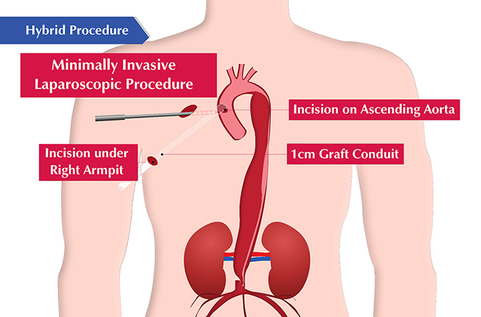
The cardio thoracic surgeons did a mini-thoracotomy using minimally invasive technique on the right side of the chest and sutured a 1 cm graft conduit in the ascending aorta and brought it out near the right armpit. This was to facilitate the endovascular team to access the aorta, in order to deploy the endovascular graft to the descending aorta.
Step 2:
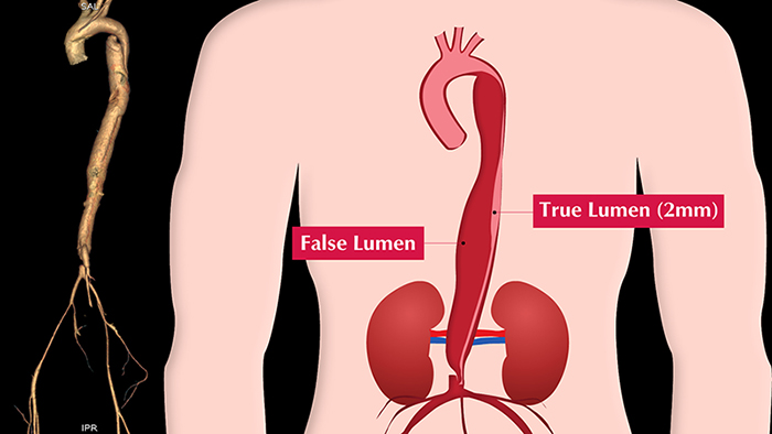
The doctors then shifted the patient to the cath lab. When the guide wire was inserted into the descending aorta the doctors were unable to proceed further as the aortic dissection was so bad – practically there was no way to differentiate between the true lumen* and the false lumen*.

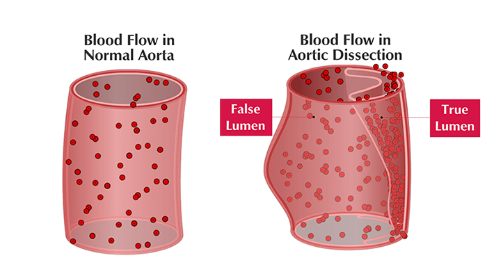
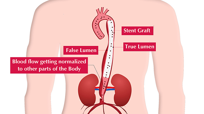
* A dissection in aorta occurs when a tear of the intima (the inner lining) allows blood to leak into the media (middle layer). This creates two passages for blood: a true lumen, which is the normal passage for blood, and a false lumen, the newly created passage.
Step 3:
The guide wire was inserted through the leg artery and passed through the 2mm true lumen and came out through the specially sutured 1 cm graft conduit under the right armpit. Later with lot of technical challenges the doctors successfully deployed a full size endovascular graft (via this 1 cm graft conduit) in to the entire descending aorta.

Post Surgery
Post OP care:
Bharath was taken off the ventilator the next day and within 10 days he was discharged home. Today Bharath has recovered very well and can walk 1 km without any hassle. Bharath is the youngest patient treated in India with such complexity.
This amazing result was achieved only because of the multi-disciplinary team approach at MIOT towards patient’s outcome and standing by our motto “Putting Patients First”
About MIOT INSTITUTE OF CARDIAC CARE
From treating life threatening heart attacks to taking care of days old blue babies, MIOT Institute of Cardiac Care has both the infrastructure and expertise to offer patients care for the entire spectrum of diseases and disorders.
The MIOT Centre for Thoracic and Cardiovascular surgery performs more than 1000 surgeries annually which includes Coronary Artery Disease, Valvular Heart disease, Diseases of Aorta, Heart failure treatment, General Thoracic surgeries, Vascular surgeries and Adult Congenital heart diseases with a success rate matching global standards.
Brief outline on Aortic Dissection
Aortic dissection occurs when there is injury to the innermost layer of the aorta (the large blood vessel of the body supplying blood to the rest of the body from the heart). Blood starts flowing between the layers of the aortic wall leading to separation of the layers within the aortic wall.
When left untreated, about 33% of those affected will die within the first 24 hours, 50% die within the first 48 hours. And 75% can die in 2 weeks’ time when the ascending aorta is involved.
Any disease which weakens the strength of the aortic wall will predispose to aortic dissection. The common risk factors are: aging (most patients are in the 40-70 years age group), atherosclerosis, hypertension, blunt trauma and deceleration injuries like those which happen in road accidents, connective tissue disorders and sometimes in pregnancy.